Several commissions, appointed during the first quarter of this century to investigate the cause of pellagra, concluded from their studies that pellagra was an infectious, contagious disease. Harris (1913) was able to inject Berkefeld filtered tissue material from pellagra victims into monkeys to cause a corresponding disease in these animals. He concluded from these experiments that a virus was present in the injected material and that it was the cause of pellagra. If the work of Harris had been followed exclusively, various strains of this "virus" might have been discovered and a vaccine, effective in experimental animals, might have been developed, as in the case of poliomyelitis. Today, as a result of unlimited research, however, we know conclusively that pellagra is not caused by a virus but rather that it is a vitamin deficiency disease. It is obvious that if the investigations of pellagra had been restricted to the virus theory, it would still be a mystery. (R. Scobey, 1952)
From J.A.M.A., June 21, 1913, vLXp1948
The
Experimental Production
Of Pellagra In The Monkey
By A
Berkefeld Filtrate Derived From Human
Lesions -- A Preliminary Note*
William H. Harris, M.D.
The inability to produce satisfactorily pellagra in animals by means of various foodstuffs (spoiled maize and other cereals) which are considered by many observers to be in some manner responsible for the production of the disease, led to investigations based on the hypothesis that pellagra is caused by a living microorganism and not by a chemical intoxicant. Therefore, it seemed logical that the causal agent would be contained in one or more of the various tissues affected, and by utilizing these, the disease could be reproduced in a susceptible animal. It was considered inadvisable to attempt injections of the whole tissue emulsions because of the heavy bacterial flora of the intestinal tract, the contaminations of the skin and the probable secondary infection of the patient dead of pellagra. These considerations together with the view that the disease in man might be due to a filterable virus occasioned the employment of filtrates from the various organs. For this purpose the skin, alimentary tract and more especially the brain and cord, since many of the characteristic symptoms of pellagra indicate disorders of this system, were filtered and utilized for animal injection.
In the spring of 1910 experiments with a Berkefeld filtrate of the infected human tissues were carried out on the monkey. These materials were selected from the fresh necropsy of a case of undoubted pellagra which presented clinically a typical picture of the disease; namely, extensive skin lesions, stomatitis, diarrhea and the various nervous manifestations. The skin lesions involved the hands, face, legs and scrotum, and were sharply defined, being of a distinct black color, dry, elevated and scaly. A complete necropsy was held within two hours after death, and the only lesions found in the gross and microscopically were those present in fatal pellagra; no concomitant disease was present. The tissues of the different parts of the central nervous system, especially the cord, portions of the skin lesions and of the alimentary tract including the nasopharyngeal mucous membrane were removed. These were mixed with equal amounts of normal saline solution, ground together in a mortar and allowed to stand in the ice-chest over night. After coarse filtration the juice was passed through a Berkefeld filter, letter N.
As the primary aim was to infect an animal with the disease, the filtrate from the tissue mixture was injected subcutaneously, intravenously and intracranially in large quantities into a full-grown, normal male Macacus rhesus (Monkey 1). After showing immediate pressure symptoms the animal recovered from the operation and remained apparently normal for many months, when he developed irregular dark patches on the face, forearms, hands, back and sides of time body. He gradually grew thin and weak and finally died with all the signs of pellagra. The length of time for the development of the disease in this animal occasioned considerable skepticism of the results and the experiment was not repeated until a similar typical fatal human case was again available. The necropsy protocol of this animal is given below.
On Dec. 2, 1912, a full-grown healthy Macacus rhesus (Monkey 2) was inoculated with large quantities of a filtrate from parts similar to those previously used, which were obtained from a fatal case of recurrent pellagra that showed on post-mortem examination no evidences of other disease. The injections were given intracranially, intravenously and subcutaneously and, aside from some immediate pressure symptoms, the animal remained apparently normal for a period of two months.
Another distinct case of fatal pellagra presented itself in February which revealed at necropsy only the lesions consistent with pellagra. The small intestine showed extensive inflammation of the mucosa. Two separate filtrates for animal inoculation were prepared from the tissues of this subject: two from the brain and cord and another from the intestinal and skin lesions. The nasopharyngeal mucous membrane was not used and only a very small amount of skin. The animal which had previously received injections of pellagra filtrate (Monkey 2) was given a second injection of equal parts of these two filtrates mixed together. Another animal of the rhesus type (Monkey 3) was injected intravenously and intracranially with the filtrate prepared from the brain and cord. These two animals were inoculated on Feb. 12, 1912.
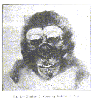
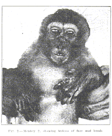
Early in May, Monkey 2, which had received two inoculations, one in December and the other in February, showed irregular patches of a copper or dusky red color about the face. On May 10 these patches were raised, considerably darkened, very dry clearly across the nose and more defined, being located across the nose and spreading in wing-like manner on either side under the eye and over the cheek. The shape of the lesion in general was somewhat that of a butterfly. At this time copper-colored lesions had appeared on the summit of the concha of both ears and at the external canthus of the eyes and also over the backs of the hands. There was a distinct symmetry in the bilateral arrangement and shape of these lesions. Subsequently the animal has become melancholic, emaciated and weakened: he sits about the cage (dimensions 8 by 10 by 15 feet). He slowly climbs instead of jumping about as previously. The lesions apparently itch since he scratches them frequently and often is seen picking off the scaly plaques. His appetite is poor. A slight diarrhea has developed and he shows considerable salivation, slobbering freely: the tip of the tongue is granular in appearance and the papulae are considerably enlarged. On May 28 the skin lesions have become almost black in color and are easily visible at a considerable distance; when viewed closely evidences of cracking and scaling are seen. At present date (June 1) the lesions look about the same except that there are deeper pigmented areas within the large plaques, producing a somewhat mosaic appearance. Along the bridge of the nose some of the scaly plaques have fallen off, one of which measures approximately 0.5 cm. The hands over the dorsal surface of the fingers and over the wrists posteriorly now present distinct dry black lesions and show, especially on the fingers, distinct scaling. The hairs over the phalanges have disappeared (see photographs). The animal is growing progressively weaker and thinner and the signs and symptoms are becoming more marked.
Monkey 3 is still in good condition and quite lively but presents about the face a few irregular, rust-colored macules, quite like the early lesions of Monkey 2, though the clinical observations on Monkey 1 are not so detailed as desired since the period of incubation was so long
that routine examinations had been discontinued, the necrospy findings are of particular interest. The animal is extremely emaciated and presents large irregular pigmented plaques on the face, forearms, and back. Those are in some areas of a chocolate color while other areas show a gray scaly surface; they are in size from approximately 2 by 3 by 3.5 cm. to 4 by 5 by 7 cm. and in other places they have merged together to form extensive patches. The lungs and the heart are normal and aside from some inflammation of the small intestine no other gross lesions are present. Microscopically, the most typical lesion is found in the sections of the skin, which show extensive hyperkeratoses of the epidermas, marked increase in the depth of the papillae and abundant pigmentation in the deeper portions of the corium where many chromatophores have wandered into the deeper strata. The cutaneous lesions are identical with the microscopic picture of the skin in human pellagra.
These experiments would indicate that pellagra may be transmitted to the monkey (Macacus rhesus) by means of a Berkefeld filtrate derived from the tissue of the human subject; at least, the animals develop all the essential clinical signs and symptoms together with the pathologic picture discerned in the disease in man. Furthermore, they suggest that the etiology of pellagra is a filterable virus or a micro-organism capable of passing through the pores of certain Berkefeld filters. The details of this work and the further experiments which have been undertaken with a view of determining the nature of this filtrate and other phases of the problem will appear in a subsequent publication.
For many suggestions during the progress of this work I wish to extend thanks to Prof. .G. W. Duval in whose laboratory the work was carried out.
*From the Laboratories of Pathology and Bacteriology, Tulane University.
A similar experiment of evidently worse quality was performed in the first experiment which established poliovirus causality, by Landsteiner and Popper, 1908. Their work is hailed as the first discovery of poliovirus causality for polio. In the 1900s it was immediately cited to reinforce already existing public health requirements which required that polio be investigated as an infective disease caused by a microorganism. A description of their historic proof is presented below by Dr. Robert W. Lovett. Lovett was to be the foremost polio authority of the era, when later, in 1921, he was called Campobello to treat Franklin D. Roosevelt, just after he was diagnosed with paralytic polio, and from then onward for several years. (See also, an article by Lovett):
ABSTRACT OF IMPORTANT LITERATURE
Bacteriology and experimental production. -- The most valuable contribution of the year toward our knowledge of the disease has been made by Landsteiner and Popper, of Vienna, who have apparently succeeded in producing the disease in monkeys by inoculation. A boy of eight died of the disease on the fourth day. The autopsy showed typical anterior poliomyelitis. In the spinal cord and cerebrospinal fluid there were no organisms to be found and cultures were sterile. Parts of the spinal cord were then emulsified in salt solution and injected into the abdominal cavity of rabbits, guinea pigs, mice and two monkeys. In the first three named no paralysis ensued and the spinal cords were normal.
The first monkey became violently ill on the sixth day and died on the eighth. He lay on the floor of his cage and his power to move his limbs was not investigated. After death changes typical of anterior poliomyeletis were found.
The second monkey was noted to have lost all power in the hind legs on the seventeenth day. No paralysis was present on the twelfth, although it may have been present before the seventeenth in some degree. He was killed on the nineteenth day and again typical pathological changes were found in the central nervous system.
From the spinal cord of this monkey inoculations were made into two other monkeys with negative results.
The conclusion of these authors is that “a so-called invisible virus, that is, one belonging to the class of the protozoa, is the cause of the disease.”
To Lovett's credit, he mentioned that others disputed the findings of Landsteiner and Popper.
Since 1908, Draper (1917) and Melnick (1947) wrote that laboratory experiments that attempt to prove poliovirus causality must use intracranial injection of sizeable quantities of poliovirus filtrate. Other methods, such as by injection intravenously with moderate quantities of filtrate, "fail to produce any disturbance." Lovett's description does not mention that Landsteiner and Popper used a filtrate, but that they used unfiltered, emulsified spinal cord matter, which, like pureed peach skin or rabbit brain cells, is since then, well known to cause fatal reactions. Landsteiner and Popper did not use intracranial injection which would have been necessary to cause paralytic polio symptoms; they injected the much more dangerous puree of spinal cord into the abdominal cavity of the monkeys. Regarding the monkey that died, "his power to move his limbs was not investigated." (!)
Monkeys, like humans, are sensitive creatures. In the laboratory environment they are often ill-treated, and malnourished (Jane S. Smith, Patenting The Sun, Polio And The Salk Vaccine (1990)). Monkeys appear to have spontaneous demyelinating diseases (Rivers (1953)).
Home
Page
* * *
(c) HARpub 1997-1999
All Rights Reserved